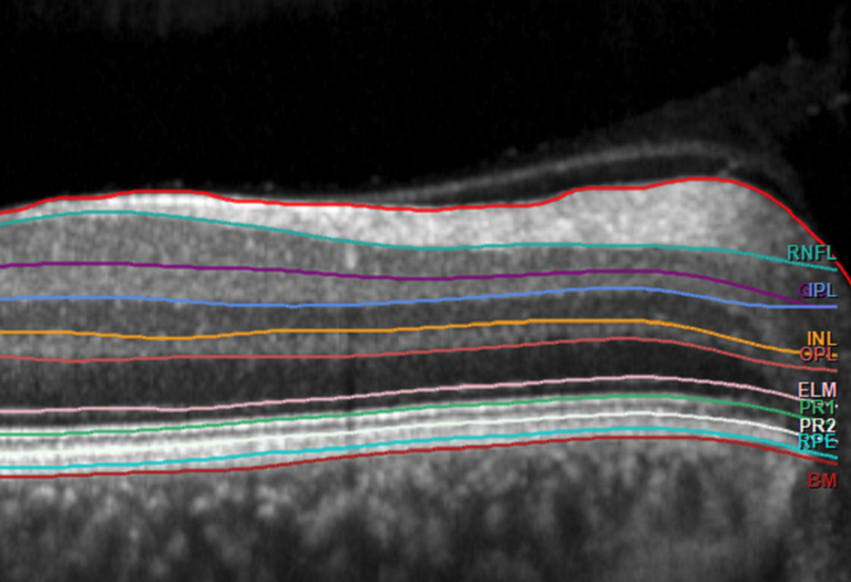
The DZNE is researching key factors for a healthy life as part of the "Rheinland Study" - a large-scale population study in the Bonn urban area. Determining biomarkers for dementia and other neurodegenerative diseases is one of the study's goals. Among other things, the focus is on the human retina: For these investigations, the DZNE is cooperating closely with the eye clinic of the UKB. "There are indications that the retina can serve as a window into the brain, so to speak. Our current results support this thesis," explains Prof. Dr. Frank G. Holz, Director of the Eye Clinic at the UKB. "Compared to previous studies, the latest technology was used here and a larger group of people was examined."
State-of-the-art imaging in the Rhineland study
In nearly 3,000 participants of the Rhineland Study between the ages of 30 and 94, the retina was examined using "spectral domain optical coherence tomography" (SD-OCT) - a procedure that allows detailed images of the retina and its various layers. In addition, the brain was measured using magnetic resonance imaging (MRI). The data were analyzed using sophisticated software algorithms. "In this way, the different layers of the retina and the different structures of the brain can be automatically recorded and their thicknesses and volumes determined. In the next step, we looked for correlations between the volume of the retina and the volume of certain brain structures," explains Dr. Dr. Matthias M. Mauschitz, assistant physician at the UKB Eye Clinic, postdoctoral scientist at the DZNE and first author of the current publication.
Thinner retinal layers with reduced brain volume
"A close correlation was shown between layers of the inner retina and the so-called white matter inside the brain," Mauschitz continued. "The thinner these retinal layers, the lower the brain white matter volume was." In contrast, parts of the outer retina were primarily associated with the gray matter of the cerebral cortex. In the so-called occipital lobe of the brain, where the visual center is located, these associations were particularly pronounced. The researchers also encountered relationships elsewhere. "Interestingly, the thickness of different retinal layers correlated closely with the volume of the hippocampus. This is an area of the brain that plays a central role in memory and is often affected in dementia," adds Prof. Dr. Dr. Robert P. Finger, senior consultant at the UKB Eye Clinic.
Progressive examination in neurodegenerative diseases?
"Images of the retina using SD-OCT are relatively easy, non-invasive and inexpensive to perform. The current results suggest that such images may be suitable as biomarkers for brain atrophy and thus could serve as additional follow-up in the case of certain neurodegenerative diseases," said Prof. Dr. Dr. Monique M. B. Breteler, director of population-based health research at the DZNE and head of the Rhineland study. "Further population-based studies as well as studies in patient groups and over a longer period of time are now needed to verify these results in a clinical setting."
Originalveröffentlichung
Retinale Schichtbewertungen als potenzielle Biomarker für Hirnatrophie in der Rheinland-Studie, Matthias M. Mauschitz et al., Scientific Reports (2022), DOI: 10.1038/s41598-022-06821-4,
URL: https://www.nature.com/articles/s41598-022-06821-4
Contact for the media:
Matthias M. Mauschitz, MD, PhD.
Bonn University Eye Hospital and Population Health Research, German Center for Neurodegenerative Diseases (DZNE)
Ernst-Abbe-Str. 2
53127 Bonn
Matthias.Mauschitz@ukbonn.de
www.ukbonn.de/augenklinik
Press contact:
Viola Röser
Deputy Press Officer at Bonn University Hospital (UKB)
Tel.: 0228 287-10469
E-mail: viola.roeser@ukbonn.de
About the German Center for Neurodegenerative Diseases (DZNE)
The DZNE is a research institution that focuses on all aspects of neurodegenerative diseases (such as Alzheimer's, Parkinson's and ALS) in order to develop new approaches to prevention, therapy and patient care. Through its ten locations, it bundles nationwide expertise within one research organization. The DZNE cooperates closely with universities, university hospitals and other institutions in Germany and abroad. It is publicly funded and a member of the Helmholtz Association. Website: www.dzne.de
About the University Hospital Bonn: The UKB cares for more than 400,000 patients per year, employs 8,300 people and has a balance sheet total of 1.3 billion euros. In addition to the more than 3,300 medical and dental students, around 600 young people are trained in other healthcare professions each year. The UKB is ranked first among university hospitals (UK) in NRW in the science ranking, has the fourth highest case mix index (case severity) in Germany and in 2020 was the only one of the 35 German university hospitals to have an increase in performance and the only positive annual balance sheet of all university hospitals in NRW.
Source: UKB Newsroom
