IMOD Workshop
Our hands-on mini-workshop on using IMOD for electron microscopy (EM) image processing and 3D reconstruction is tailored for two researchers - ideally with active volume EM projects. Participants will work on a curated 3D EM dataset to learn fundamental segmentation workflows.
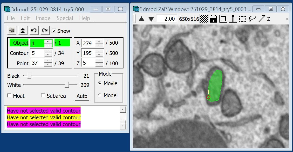
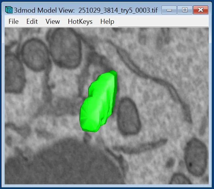
During the session, you will explore visualization, manual segmentation, and 3D model generation within IMOD. Resulting models can be exported for downstream segmentation refinement or quantitative analysis pipelines. The emphasis is on understanding the core principles of 3D EM data handling in IMOD and gaining the practical confidence to process real datasets independently.
As seats are very limited (only two), we ask you to submit a brief application describing your motivation and how to implement IMOD in your research.
During the workshop, participants will work with a FIB-SEM dataset generated by the Microscopy Core Facility to manually segment mitochondria. The objective is to introduce the core functionalities of IMOD and to provide structured, hands-on experience in navigating, annotating, and modeling 3D EM volumes.
You can find many details about ilastik on its website: https://bio3d.colorado.edu/imod/
The Core Facilities thank the German Research Foundation for continuous support.