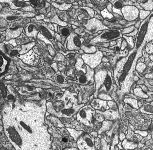
Volume Electron Microscopy / FIB-SEM
Our core facility offers volume electron microscopy using FIB-SEM (Focused Ion Beam Scanning Electron Microscopy) for high-resolution 3D reconstruction of ultrastructural features. This technique combines precise ion beam milling with serial SEM imaging, allowing detailed visualization of internal structures across volumes typically in the range of a single cell (e.g., ~10 × 10 × 10 µm³). FIB-SEM is especially suited for exploring the organization of subcellular components in three dimensions at nanometer resolution. Automated acquisition workflows ensure consistent data quality and enable targeted volume analysis with high spatial accuracy.
If you are interested, please contact us.
The Core Facilities thank the German Research Foundation for continuous support.