Instruments
The Microscopy Core Facility (MCF) provides access to a wide range of state-of-the-art light and electron microscopy systems for biomedical research. Our instruments support high-resolution imaging, live-cell microscopy, super-resolution techniques, and correlative light and electron microscopy (CLEM). All systems are maintained, regularly updated, and embedded in a quality-controlled service environment. Researchers receive hands-on training, project support, and access to specialized workflows tailored to their scientific questions.
Whether you're studying living cells, tissue architecture, or subcellular ultrastructure — the MCF offers the right tools and expertise to advance your imaging needs. Feel free to contact us and we will discuss your projects with you to find the most appropriate imaging technology for your needs.
-
 © MCF
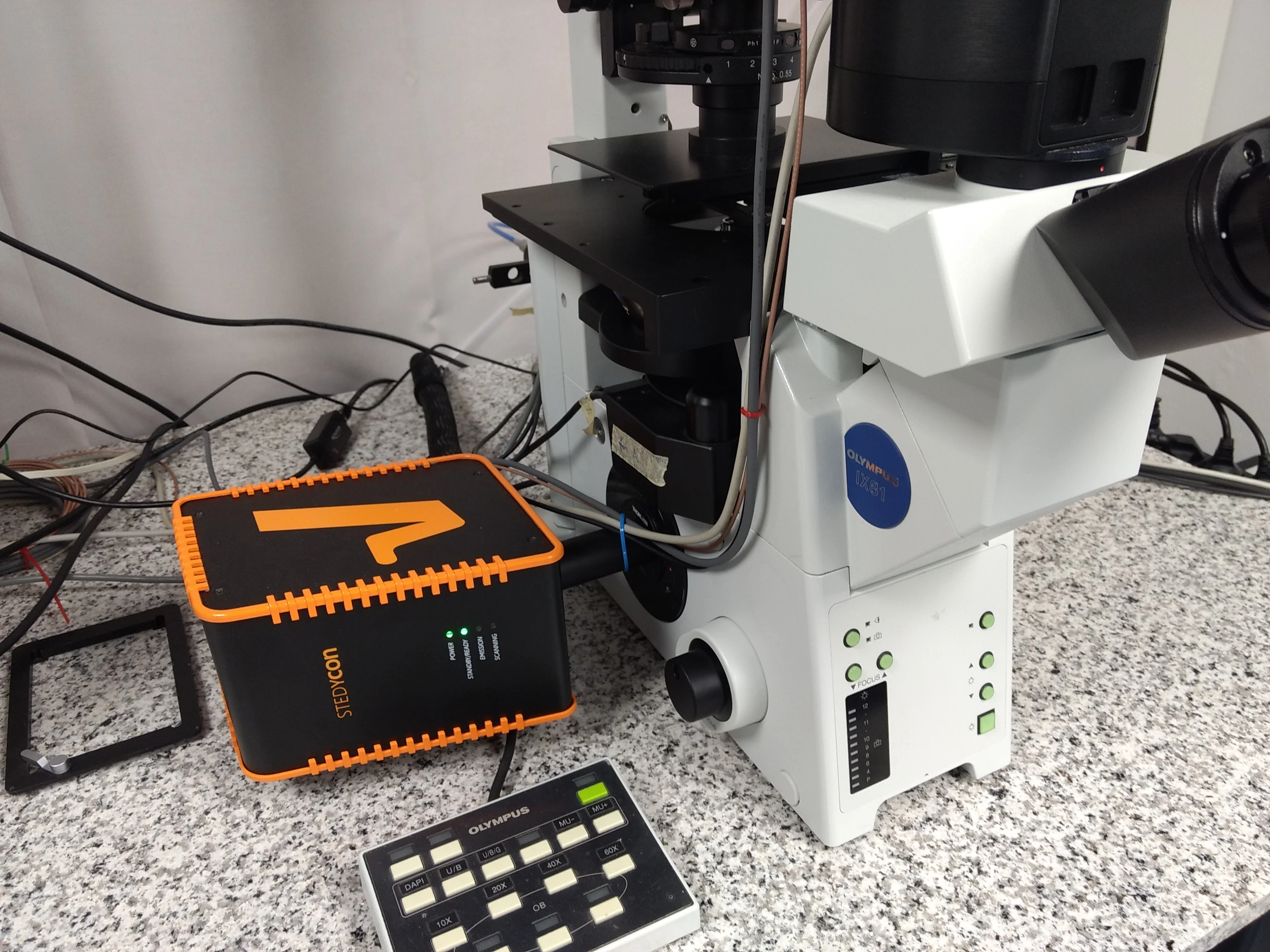
© MCFAbberior STEDYCON
The Abberior STEDYCON is mounted on our Olympus IX81 microscope. The STEDYCON module contains four exitation laser lines and an additional depletion laser (750nm) for STED superresolution imaging.
-
 © MCF
© MCFLeica SP8 Lightning
Point-scanning confocal microscope for imaging fixed and live samples with high-resolution.
-
 © Rolf Müller
Leica Stellaris 8
Point-scanning confocal microscope for imaging fixed and live samples with high-resolution.
-
 © MCF
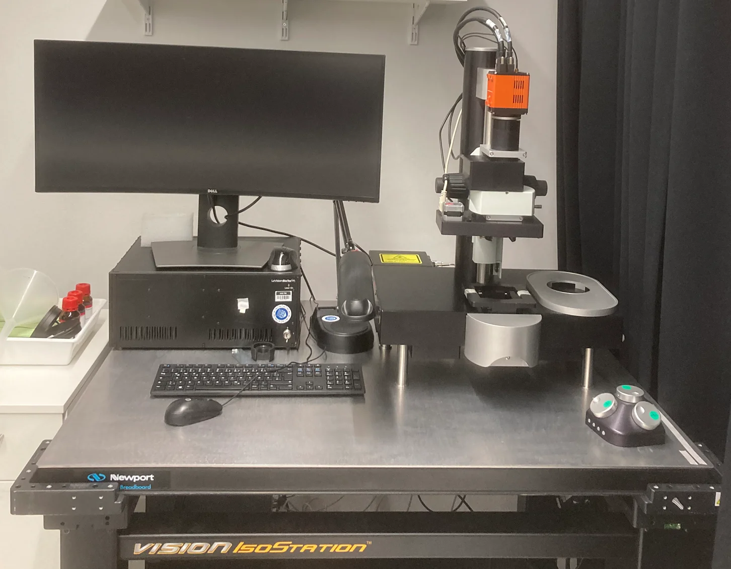
© MCFMiltenyi LaVision Ultramicroscope II
The Miltenyi LaVision Ultramicroscope II is a lightsheet microscope that can be used for fast 3D imaging of large cleared specimens.
-
© MCF
Olympus IX81
The Olympus IX81 is a motorized inverted fluorescence microscope. It supports transmitted light and fluorescence imaging with multiple filter configurations, allowing observation of fixed specimens. With the attached Abberrior STEDYCON module, also superresolution imaging is possible. The motorized stage and focus control enable automated image acquisition for multi-position experiments. Although an older model, the IX81 remains a dependable system for everyday microscopy tasks.
-
 © MCF
© MCFVisitron Visicope
The Visitron VisiScope system with Homogenizer is a high-end fluorescence imaging platform optimized for live-cell imaging and larger volume acquisitions. Integrated with a homogenizer for uniform illumination across the field of view, the system ensures consistent excitation intensity. The microscope supports widefield and spinning disk confocal imaging with sensitive sCMOS cameras at high speed.
-
© Volker Lannert/Uni Bonn
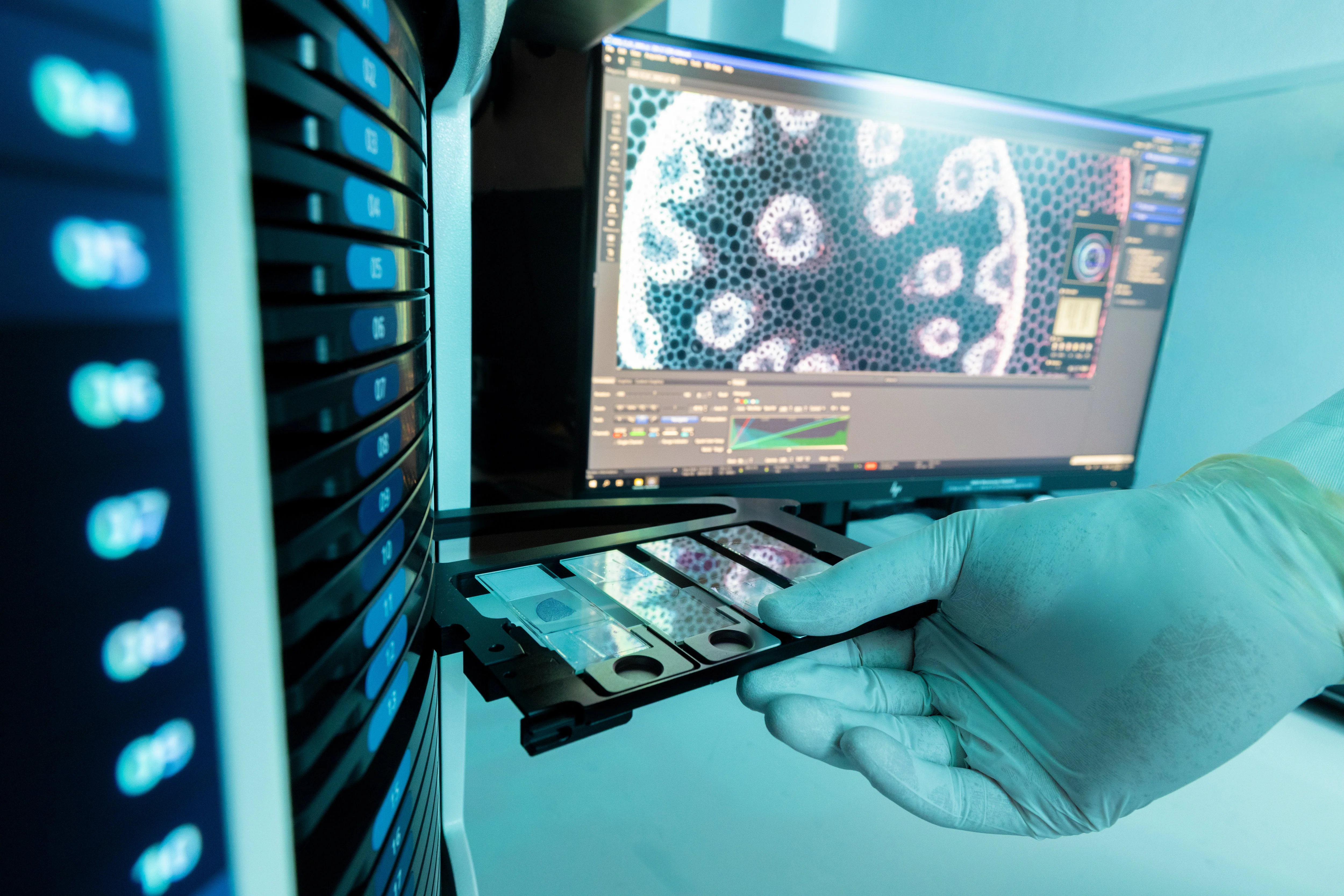
Zeiss Axioscan.Z1
The Zeiss Axioscan.Z1 is a high-performance, fully automated slide scanner designed for high-throughput whole slide imaging. It supports brightfield and fluorescence widefield modalities, offering up to 100-slide capacity for uninterrupted batch scanning. Its flexible configuration supports multiple fluorescent channels and advanced autofocus routines, making it ideal for digital pathology, research, and education. The intuitive software interface ensures seamless workflow integration, from slide loading to data management and export.
-
 © MCF
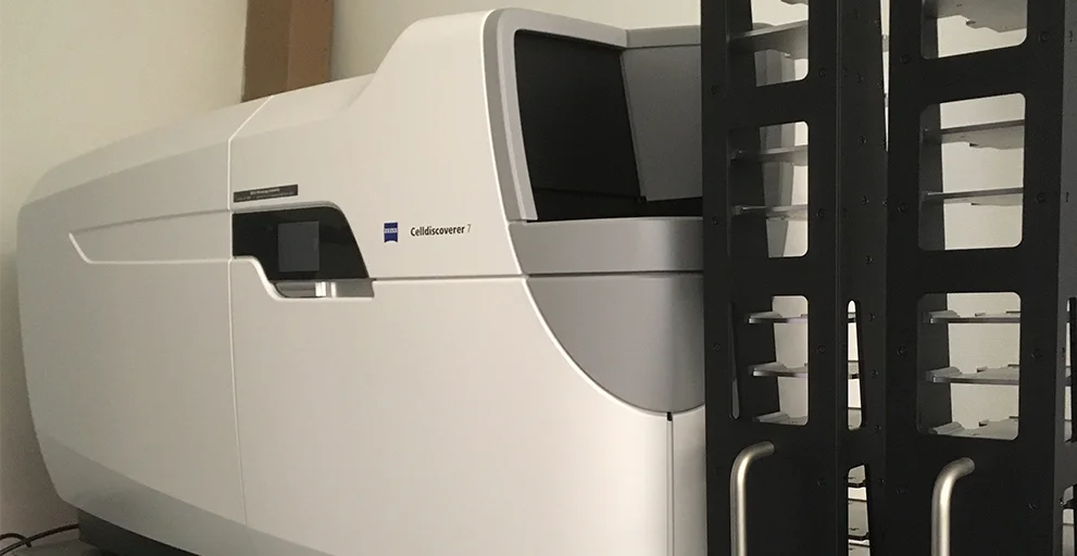
© MCFZeiss CellDiscoverer 7
High-content screen widefield fluorescence microscope for automated imaging of several multi-well plates, equipped with plate loader robotics. The incubation chamber allows for temperature and CO2 control to provide optimal conditions for live cell imaging.
-
© Volker Lannert/Uni Bonn
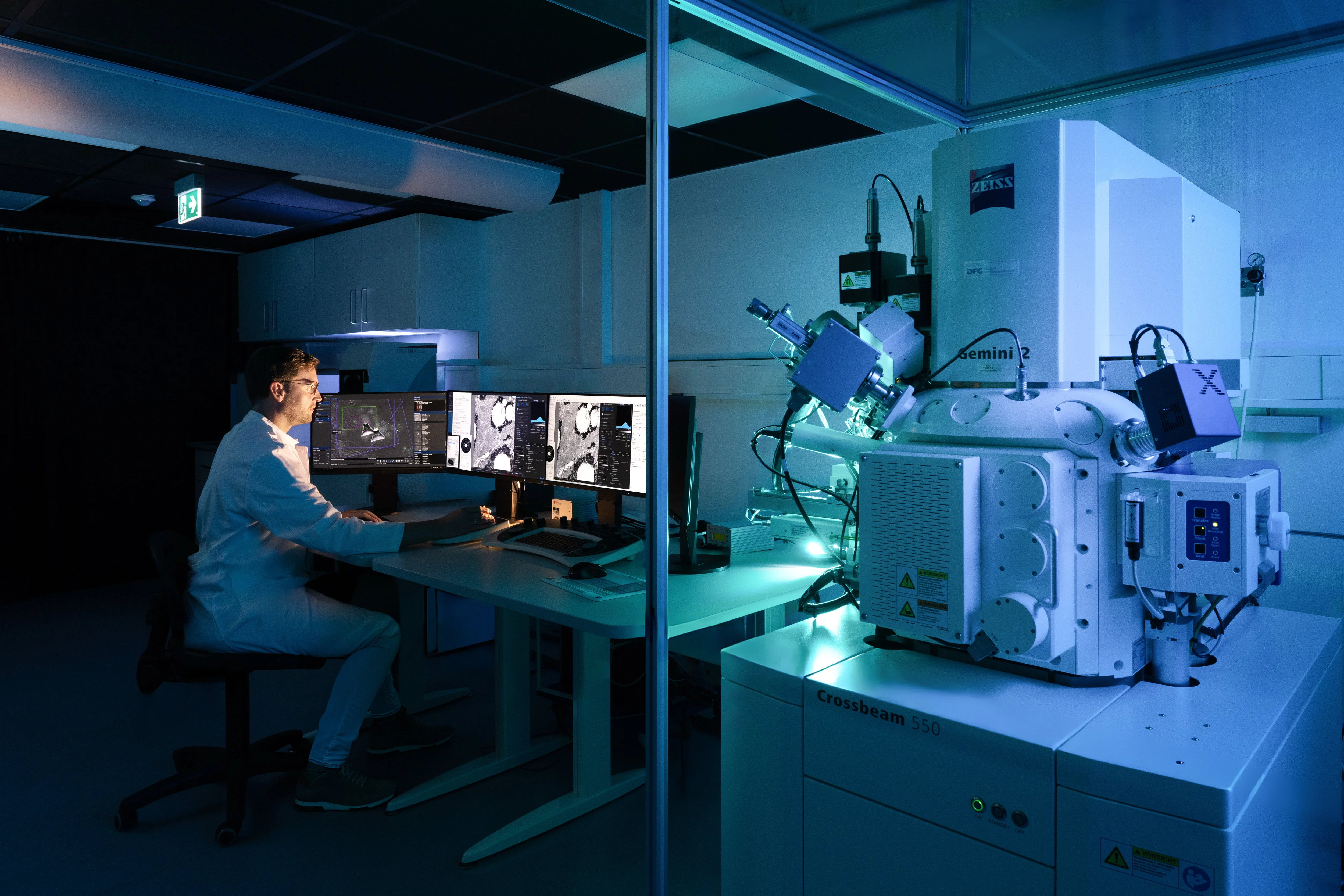
Zeiss Crossbeam 550
High-resolution FIB-SEM system for 2D and 3D imaging of biological and material samples. Equipped with SE, BSE, EsB, Inlens, and STEM detectors, it enables flexible contrast modes for surface, compositional, ultrastructural, and transmission imaging. The integrated Capella FIB column allows for precise milling and volume imaging at nanometer resolution. Ideal for volume electron microscopy, transmission analysis of thin samples, and correlative workflows.
-
© Volker Lannert/Uni Bonn
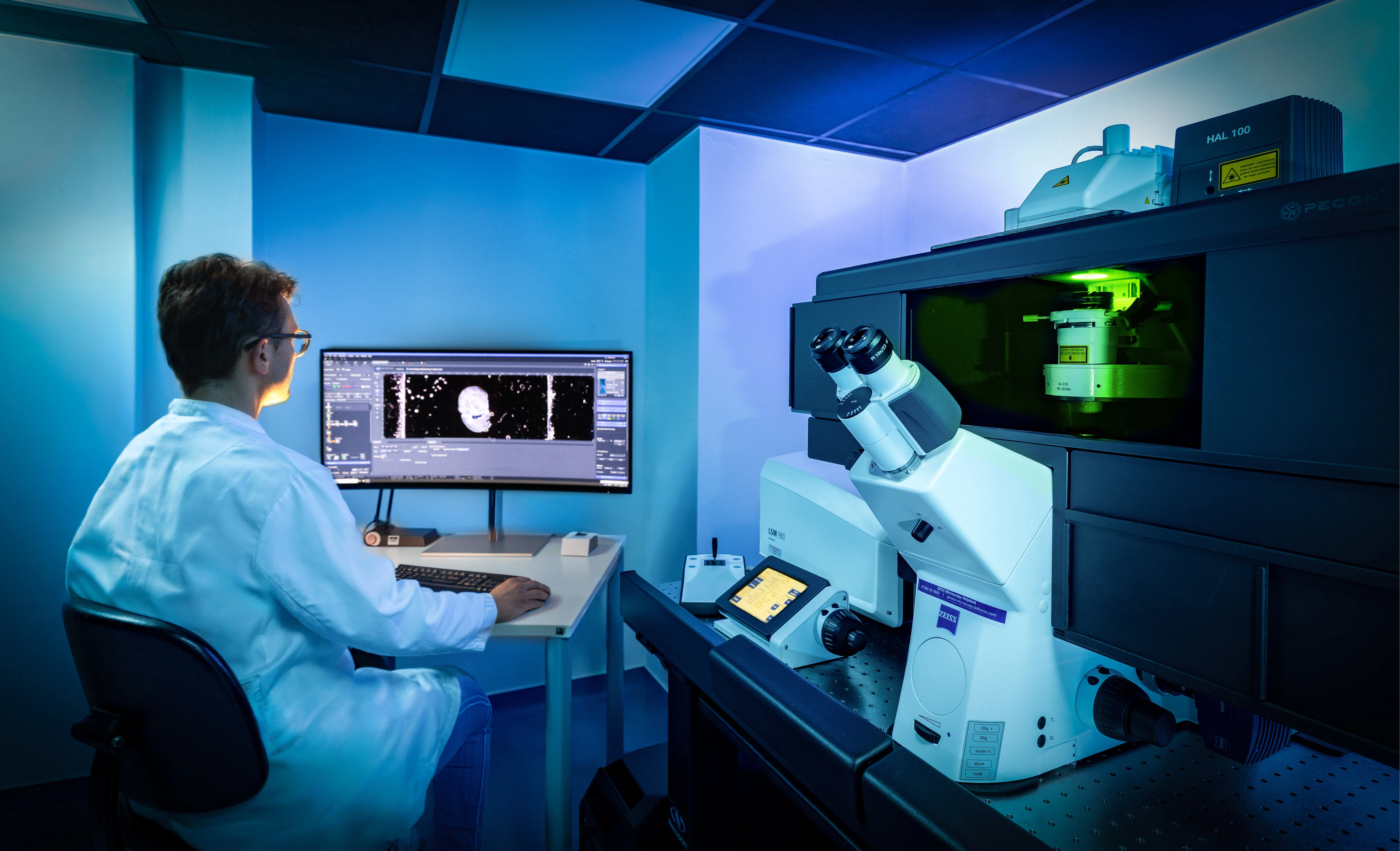
Zeiss LSM980 with Airyscan 2
The Zeiss LSM 980 with Airyscan 2 is a state-of-the-art confocal laser scanning microscope designed for cutting-edge imaging in biomedical and life sciences research. Featuring the advanced Airyscan 2 detector, the system delivers super-resolution imaging beyond the diffraction limit while maintaining high sensitivity and speed. Its unique multiplexed detection enables fast, gentle imaging of live samples with enhanced resolution and signal-to-noise ratio. The LSM 980 supports a wide spectral range, seamless multi-channel acquisition, and precise Z-stack and 3D imaging, making it ideal for complex, dynamic biological systems. Combined with intuitive ZEN software and seamless integration of environmental control and automation, the LSM 980 provides unparalleled flexibility and performance for both fixed and live-cell imaging.
-
 © Rolf Müller
Zeiss Observer.Z1
Widefield fluorescence microscope for live cell imaging and fixed samples. Dual cam supports acquisition of two channels in parallel. The incubation chamber allows for temperature and CO2 control to provide optimal conditions for live cell imaging.
The Core Facilities thank the German Research Foundation for continuous support.